Microscopy

Field Emission Scanning Electron Microscope (FE-SEM)
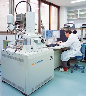
| The field emission scanning electron microscope enables the clear observation of the minute surface irregularity of solid samples beyond the reach of an optical microscope by scanning the surface with narrow electron beams after rendering it electrically conductive through the deposition of metal or carbon and by detecting and measuring the intensity of reflected electrons and secondary electrons. | ||||||||
| Model | ||||||||
|---|---|---|---|---|---|---|---|---|
| JSM-6500F | ||||||||
| Manufacture | ||||||||
| JEOL Ltd. | ||||||||
| Operating condition | ||||||||
|
||||||||
| Lab. | ||||||||
| X-Ray & SEM |
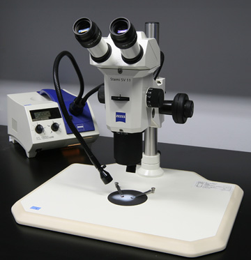
Stereomicroscope
| - | ||||||||||||||||||||||||||
| Model | ||||||||||||||||||||||||||
|---|---|---|---|---|---|---|---|---|---|---|---|---|---|---|---|---|---|---|---|---|---|---|---|---|---|---|
| Stemi SV11 (SV11-R), Stemi SV6, Stemi 2000 | ||||||||||||||||||||||||||
| Manufacture | ||||||||||||||||||||||||||
| Carl Zeiss MicroImaging Japan | ||||||||||||||||||||||||||
| Operating condition | ||||||||||||||||||||||||||
Stemi SV11
Stemi SV6
Stemi 2000
|
||||||||||||||||||||||||||
| Lab. | ||||||||||||||||||||||||||
| X-Ray & SEM |
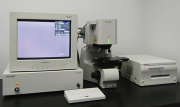
Color laser microscope
| - | ||||||||||||||||
| Model | ||||||||||||||||
|---|---|---|---|---|---|---|---|---|---|---|---|---|---|---|---|---|
| VK-8550 | ||||||||||||||||
| Manufacture | ||||||||||||||||
| Keyence Corporation | ||||||||||||||||
| Operating condition | ||||||||||||||||
VK-8550 ÁE1 microscope
|
||||||||||||||||
| Lab. | ||||||||||||||||
| X-Ray & SEM |
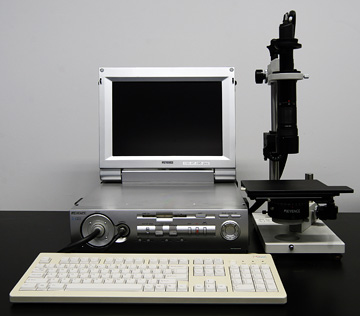
Digital microscope
| - | ||||||
| Model | ||||||
|---|---|---|---|---|---|---|
| VH-8000 | ||||||
| Manufacture | ||||||
| Keyence Corporation | ||||||
| Operating condition | ||||||
|
Keyence Digital HF Microscope System
Transmission microscope |
||||||
| Lab. | ||||||
| X-Ray & SEM |
CCD Color Digital Camera System
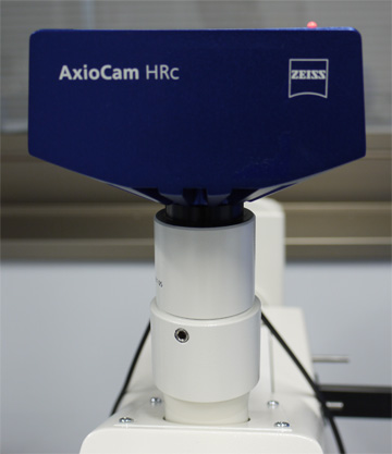
| The high-resolution camera for digital documentation can be attachable with inverted fluorescence microscope, polarization microscope and universal photo microscope | ||||||||||
| Model | ||||||||||
|---|---|---|---|---|---|---|---|---|---|---|
| AxioCam HR | ||||||||||
| Manufacture | ||||||||||
| Carl Zeiss MicroImaging Japan | ||||||||||
| Operating condition | ||||||||||
CCD Color Digital Camera System: Zeiss AxioCam HR
|
||||||||||
| Lab. | ||||||||||
| X-Ray & SEM |
Film camera
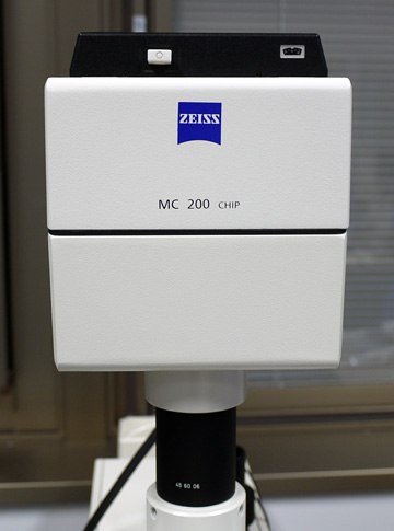
| Film camera for analogue documentation can be attachable with inverted fluorescence microscope, polarization microscope and universal photo microscope | ||||||
| Model | ||||||
|---|---|---|---|---|---|---|
| MC200 CHIP
|
||||||
| Manufacture | ||||||
| Carl Zeiss MicroImaging Japan | ||||||
| Operating condition | ||||||
Zeiss MC200 CHIP
|
||||||
| Lab. | ||||||
| X-Ray & SEM |
Universal Photo Microscope
| - | ||||||||||||||
| Model | ||||||||||||||
|---|---|---|---|---|---|---|---|---|---|---|---|---|---|---|
| Axiophoto2 (PH2-FL/Ph/DIC) | ||||||||||||||
| Manufacture | ||||||||||||||
| Carl Zeiss MicroImaging Japan | ||||||||||||||
| Operating condition | ||||||||||||||
|
||||||||||||||
| Lab. | ||||||||||||||
| X-Ray & SEM |
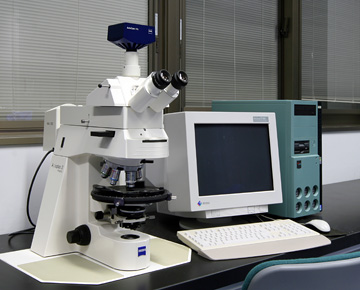
Inverted Microscope
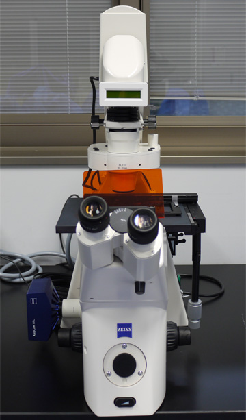
| - | ||||||||||||
| Model | ||||||||||||
|---|---|---|---|---|---|---|---|---|---|---|---|---|
| Axiovert (V200M-FL/PH/DIC) + AxioCam HR | ||||||||||||
| Manufacture | ||||||||||||
| Carl Zeiss MicroImaging Japan | ||||||||||||
| Operating condition | ||||||||||||
Axiovert V200M ÁE2 microscopes
|
||||||||||||
| Lab. | ||||||||||||
| X-Ray & SEM |
Polarizing Microscope

| - | ||||||||||||||||||||||||
| Model | ||||||||||||||||||||||||
|---|---|---|---|---|---|---|---|---|---|---|---|---|---|---|---|---|---|---|---|---|---|---|---|---|
| Axioskop 40 A Pol, Axioplan2 Imaging Pol (P2IPOL-T/R2), Axiolab Pol (LABPOL-T/RS), Axiolab Pol (LABPOL-T1), | ||||||||||||||||||||||||
| Manufacture | ||||||||||||||||||||||||
| Carl Zeiss MicroImaging Japan | ||||||||||||||||||||||||
| Operating condition | ||||||||||||||||||||||||
Axioskop 40 A Pol
Axioplan2 Imaging Pol
|
||||||||||||||||||||||||
| Lab. | ||||||||||||||||||||||||
| X-Ray & SEM |